
Click for full-size.


An Atlas of the Fertilization and Karyokinesis of the Ovum
by Wilson, E.B. and Leaming, E
- Used
- first
- Condition
- See description
- Seller
-
North Garden, Virginia, United States
Payment Methods Accepted
About This Item
New York: Columbia University Press, Macmillan and Co., 1895. First edition.
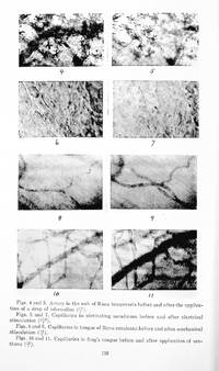
1895 FOLIO ATLAS OF FIRST PUBLISHED PHOTOGRAPHS OF CHROMOSOMAL DIVISION BY LEADING AMERICAN CELL BIOLOGIST.
11x13 inch folio hardcover, dark blue cloth binding, gilt title, "Columbia University Biological Series" to cover, gilt title to spine, back cover blindstamped with Macmillan colophon, bookplate of University of London Library and "withdrawn" handstamp to front paste-down--no other marks. Wear to cover corners, text and plates clean and bright: very good in custom archival mylar cover. FROM THE PREFACE: "It is the object of this work to place before teachers and students of biology a series of figures, photographed directly from nature, to illustrate some of the principal phenomena in the fertilization and early development of the animal ovum. In no branch of biological inquiry has knowledge advanced of late with such rapid strides as in the new science of cytology, which deals with the internal phenomena of cell-life. Within the past two decades this science has brought forward discoveries relating to the fertilization of the egg and the closely related subjects of cell-division and karyokinesis that have called forth, on the part of Weismann and others, some of the most important and suggestive discussions of the post-Darwinian biology. These discoveries must in some measure be dealt with by every modern text-book of morphology or physiology, yet they belong to a region of observation inaccessible to the general reader or student, since it can only be approached by means of a refined histological technique applied to special objects not ordinarily available for practical study or demonstration. A knowledge of the subject must, therefore, as a rule, be acquired from text-books in which drawings are made to take the place of the real object. The plates of this atlas are reproduced from photographs of the eggs of the sea-urchin, Toxopneustes variegatus, hg. procured at Beaufort, N.C. The eggs, carefully selected from ripe females, were artificially fertilized in sea-water, and preserved at regular intervals. The eggs or this species have one great advantage of being devoid of pigment and very transparent, so that nuclei, asters, and spindles can be clearly seen and their general history followed in life." Cited by J Maienschein in Embryos Under the Microscope (2014): "In the United States, Edmund Beecher Wilson's works in 1895 and 1896 became the classic English-language discussions of the role of the cell. Wilson had been one of the first to study at the new Johns Hopkins University biology department, the formation of which had followed the inspiration of leading graduate educational institutions in Germany and England. Wilson came from a farm background in Ohio, and the more he learned about biology, the more he became fascinated by cells and developing organisms. After writing the first major introductory textbook on general biology with his fellow Johns Hopkins graduate William Sedgwick, Wilson turned to a detailed study of cells. Though his first jobs after receiving his doctorate included a series of short stays at Williams College, then the Massachusetts Institute of Technology, and then the excellent women's college Bryn Mawr, in 1891 he moved to Columbia University in New York where he remained for the rest of his career. In New York, Wilson partnered with a photographer to record exactly what happens in the earliest stages of development, the early cell divisions. His first studies looked at the polychaete Nereis worms, which he collected at the Marine Biological Laboratory (MBL) in Woods Hole, Massachusetts. Wilson must have gone out on the dock of the Eel Pond to the laboratory, shone a light on the water to mimic the full moon, and caused the worms to rise to the surface toward the light and begin their mating dance. Wilson would have gathered them up in his collecting net, taken the jar back to the laboratory, collected the fertilized eggs, and put them under the microscope. He would keep watch throughout the night to see every cell division and then observe and record every step of the epigenetic process. The fertilized egg cells that Wilson watched so closely began dividing, gradually into two cells, then four, then eight and so on. As they divided, the nucleus did a wonderful dance, complete with complex spindle fibers, asters, and other apparatuses to help the division along. In later works, Wilson represented in diagrams or line drawings what he thought was important about what he saw. For his Atlas of Fertilization and Karyokinesis of the Ovum in 1895 (offered here), he photographed the details of cell division so that everyone else could also observe precisely what he had been seeing. The photographs showed in detail what occurs in the cytoplasm and the nucleus as well as all the other small structures that were not very well understood at the time. Such photography was not easy, and capturing the details of cell division can remain a challenge even today with improved technology. Today, photography and even capturing observations on video or with increasingly sophisticated digital technologies has become a routine part of cell biology, but in Wilson's day, it was rare to take photographs and expensive to publish them; a researcher made a considerable commitment. It is worth reflecting on how often today we just snap a photograph without thinking much about the process or about what selections we make in choosing one image or video over another. Wilson thought through his selections very carefully to share his observations with his readers, and it is fascinating to see what he considered important enough to record. In 1895, Wilson enlisted the photographic skills of Edward Leaming, Columbia University supported what must have been a highly expensive publication, and the large format Atlas appeared in print, These photographs were magnificently detailed and show the steps of cell division-we can observe the way that mitotic apparatus works as the cells divide and as the chromosomes divide. Wilson gave us the first published photographs of chromosomal division. He assumed that every reader would see what he saw in the photos, so rather than extracting or abstracting the parts he considered most important, he presented the "complete facts" to the public through the images.
Reviews
(Log in or Create an Account first!)
Details
- Bookseller
- Biomed Rare Books
 (US)
(US)
- Bookseller's Inventory #
- 1366
- Title
- An Atlas of the Fertilization and Karyokinesis of the Ovum
- Author
- Wilson, E.B. and Leaming, E
- Format/Binding
- Cloth binding
- Book Condition
- Used
- Quantity Available
- 1
- Edition
- First edition
- Publisher
- Columbia University Press, Macmillan and Co.
- Place of Publication
- New York
- Date Published
- 1895
- Weight
- 0.00 lbs
- Keywords
- atlas; biology; cell biology; cytology; photography; embryology; America
Terms of Sale
Biomed Rare Books
All items subject to prior sale. Orders are carefully packaged prior to shipping. Shipping charges are based on cost, and varies by destination, carrier and mail class. For heavy volumes and for all international shipments (outside the United States), please inquire shipping costs before placing your order (info@biomedrarebooks.com).
30 day return guarantee, with full refund including original shipping costs for up to 30 days after delivery if an item arrives misdescribed or damaged.
30 day return guarantee, with full refund including original shipping costs for up to 30 days after delivery if an item arrives misdescribed or damaged.
About the Seller
Biomed Rare Books
Biblio member since 2021
North Garden, Virginia
About Biomed Rare Books
I established BioMed Rare Books in 2015 as an internet-based bookshop specializing in rare and antiquarian books and papers in medicine and the life sciences. I have been collecting and studying printed works in these fields for many years, an activity that has enhanced and informed my practice of medicine and my own biological research.
Glossary
Some terminology that may be used in this description includes:
- Cloth
- "Cloth-bound" generally refers to a hardcover book with cloth covering the outside of the book covers. The cloth is stretched...
- Bookplate
- Highly sought after by some collectors, a book plate is an inscribed or decorative device that identifies the owner, or former...
- Gilt
- The decorative application of gold or gold coloring to a portion of a book on the spine, edges of the text block, or an inlay in...
- First Edition
- In book collecting, the first edition is the earliest published form of a book. A book may have more than one first edition in...
- Colophon
- The colophon contains information about a book's publisher, the typesetting, printer, and possibly even includes a printer's...
- Folio
- A folio usually indicates a large book size of 15" in height or larger when used in the context of a book description. Further,...
- New
- A new book is a book previously not circulated to a buyer. Although a new book is typically free of any faults or defects, "new"...
- Spine
- The outer portion of a book which covers the actual binding. The spine usually faces outward when a book is placed on a shelf....
Frequently asked questions
This Book’s Categories

Collectible Christmas Cards
Many of us love giving and receiving holiday cards - but did you know many of them are highly collectible? Whether signed by famous authors or just beautiful to look at, these collectible Christmas cards are sure to put a twinkle in your eye.
Collecting The Beat Generation
The Beat Generation was born out of WWII, and it still continues to exert considerable influence on today’s literary scene. Biblio sellers have a fantastic collection of Beat Generation books and ephemera for browsing.
Also Recommended
-

-

-
-
-

Save 10% on every purchase!
Join the Bibliophiles’ Club and start saving 10% on every book.
$29.95 / Year